Enquiry

Glass slide scanning facility

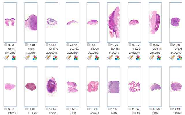
Digiscan is the best provider of slide scanning facility in India because of high resolution and excellent quality of images, competitive price and short delivery time.
Slide Scanning is carried out at 20x, 40x, 80x as per requirement using high resolution objectives to provide perfect digital images. Scanning of Histopathology, Cytology, Hematology, IHC and TMA slides is carried out.

Shape
Thumb

What is the requirement of Scanning Facility?

  • For creation of Departmental Digital slide libraries and for maintenance of permanent digital slide records of patients since cataloguing and retrieval of the digital slides is easier as compared to retrieval of glass slide.
  • For organising online conferences and slide seminars, symposia, CMEs by medical and dental faculty by digitizing cases and hosting them on server for delegates.
  • Scanning is also required for conducting EQAS
  • For Patients seeking online second opinion on their slides
  • Scanning slides of research scientists for AI diagnostics for improvement in healthcare and their other specific projects.
  • Digiscan is providing facility for scanning slides for scientific presentations for those who do not have slide scanners.
Thumb

Digiscan's Contribution to AI-Driven Cancer Diagnosis and Treatment

  • There has been a quantum jump in the number of AI companies because of the vast number of cancer patients all over the world. AI tools are being used for diagnostics as well as for treatment planning.Large number of cases of a particular cancer with its benign and normal counterpart are scanned for analysis and this creates the database for that cancer.
  • Common Cancers with Large database include CA Breast, CA Colon, CA Prostate, CA Ovary, CA Endometrium , Oral Dysplasias, Oral Carcinomas and Lymphnode metastasis. Digiscan has a large database of whole slide images in its digital library and is helping the AI companies by providing the database.
  • Similarly large database of cervical cytology ( normal and malignant) of conventional as well as LBC smears of high resolution digital whole slide images is available and is provided to AI companies who are engaged in development of software to diagnose cervical cancer using deep machine learning.
  • High Resolution WSIs Images of pathology with annotations and labelling are provided if required. WSIs of IHC markers are provided as per Research Protocol.
  • All our images are scanned and verified by a highly qualified team of Research pathologist.

Shape

Note

Kindly note that the material for scanning will have to be delivered to the scanning facility for scanning at discussed rates.

Scanning facility is provided to anatomy and pathology departments of medical and paramedical colleges for creation of departmental libraries and to research institutes for development of AI Diagnostics

Contact Us